








 Contact Platform Support
 Help Center
Home > Instruments & Meters > Test Instrument > Test Machine > Seamaty SV8 Portable Pregnancy Veterinary Color Doppler Ultrasound Machine & Portable Ultra Sound Machine
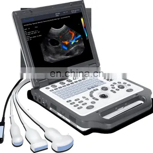
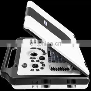
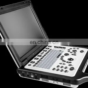
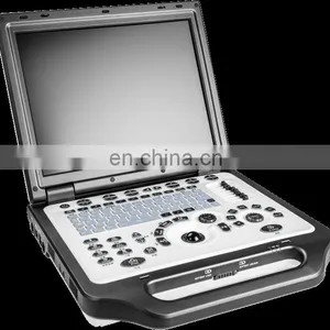
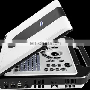
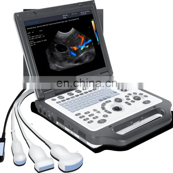
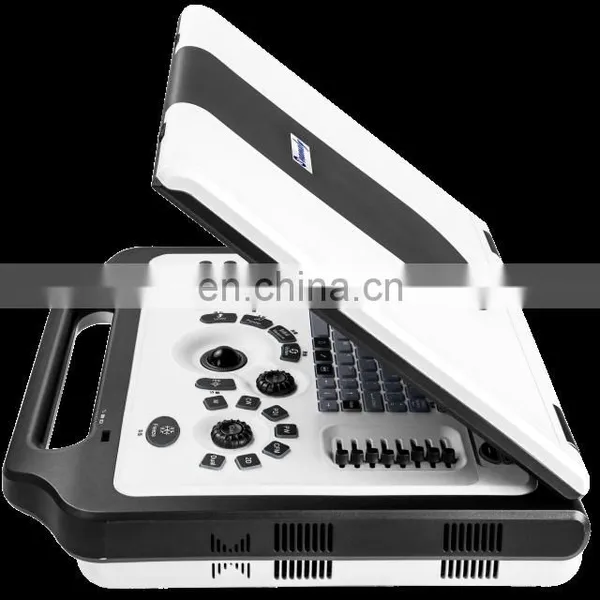
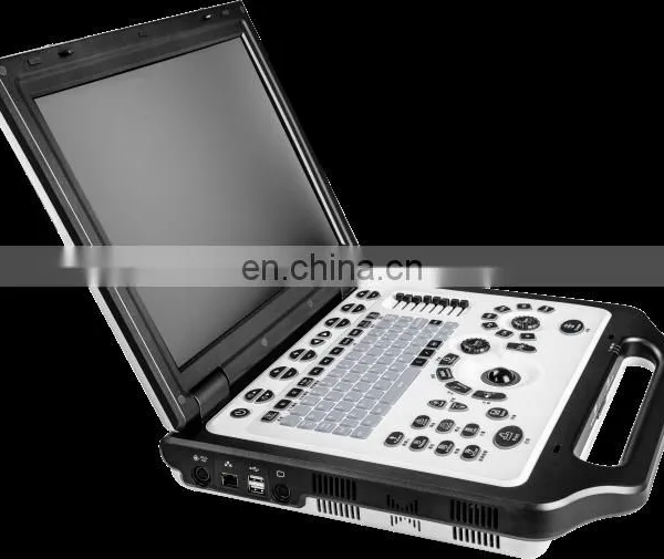
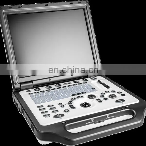
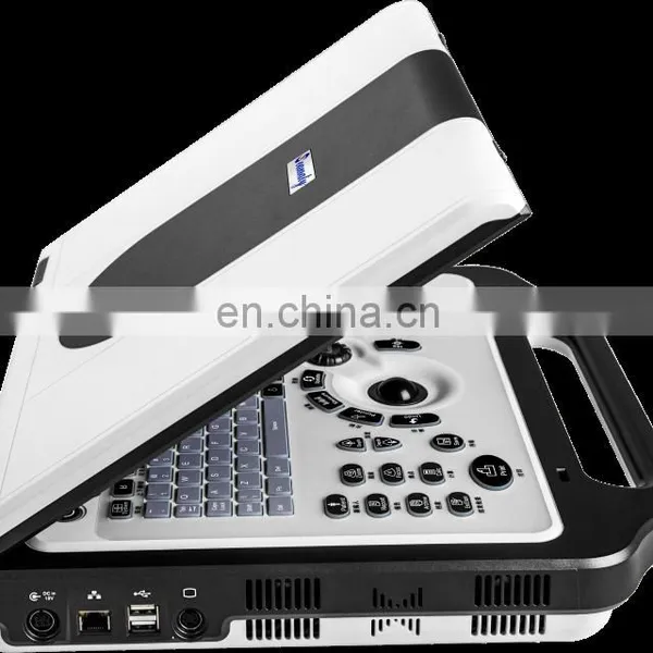
Seamaty SV8 Portable Pregnancy Veterinary Color Doppler Ultrasound Machine & Portable Ultra Sound Machine
Negotiable
MOQ: 1 set (Price negotiable depending on order volume and customization)
Key Specifications
Get Latest Price
Business Type:
Distributor/Wholesaler
Year Established:
2001
Annual Export Value:
US$5 Million - US$10 Million
Payment & Shipping
Payment Methods:

Port of Shipment:
China
Delivery Detail:
Delivery time depends on order quantity.
Related Products
-
 Cheap Portable Veterinary Ultrasound Machine With Rectal ProbesNegotiableMOQ: 1
 -
SV8 Portable Animals Ultrasound/ Veterinary Ultrasound Machine/vet Handheld Ultrasound ScannerNegotiableMOQ: 1
-
Veterinary Ultrasound Testing Equipment Portable Veterinary B UltrasoundNegotiableMOQ: 1
-
Veterinary Ultrasound Scanner Portable Color 3d Ultrasound Machine Electric Ce Mobile Trolley or Carrying Bag 24 15 LCD MonitorNegotiableMOQ: 1
-
Best Quality 2d Color Portable Veterinary Ultrasound For AnimalNegotiableMOQ: 1
Products Description
Features |
||||||||
◆ Full digital beam-former, Digital dynamic focusing, Digital variable aperture and dynamic apodizer, 32 A/D sampling channels are included for receiving and launching channels. ◆ High-density probe and wider frequency band optimize image quality of far and near field.
◆ High resolution LCD monitor, flicker-free, which can reduce the visual fatigue of the operator. ◆ The ergonomic design silicon keyboard with backlit, which is more suitable for the user. ◆ Regulated voltage power is adopted, strong suitability. |
||||||||
Standard Configuration |
SV8 Main Unit + Micro Convex / Linear Probe + Battery+DICOM 3.0 |
||||||
Display |
15“High Resolution LCD Display |
||||||
Connector |
1 Probe Connector, 3 probes connector extension optional |
||||||
Weight |
6.0kg |
||||||
Optional Configuration |
Mobile Trolley or Carrying Bag |
||||||
Languages |
English, Italian, Portuguese, Russian, Spanish, Deutsch, French |
||||||
Display Mode |
B, 2B, 4B, M, B+M, CFM, PDI, B+PW, B+CFM+PW, B+PDI+PW |
||||||
Type of Focusing |
Dynamic focusing, acoustic lens focusing, launching multipoint focusing |
||||||
Pre-processing |
low noise preamplifier, TGC, filtering, frame average, line average |
||||||
Character |
Date, time, owner name, animal name, device name, user name etc, user-defined annotation table, arrowhead and body marks Measurement Software:Software packages of animal abdomen, Obstetrics, Urology, Cardiology, and small parts; directly form diagnostic report based on measurement results. |
||||||
Body Mark |
37 body marks with probe location |
||||||
Digital Scan Converter |
628 440 24 bits |
||||||
Size |
78 mm(H)×360 mm(W)× 370 mm(L) |
||||||
◆ 1 probe connector, which can be expand to 3 connectors by the probe expansion module.15〞 high-resolution color LCD monitor. ◆ Equipped with more than 4 beam-former function
◆ Equipped with THI (Tissue Harmonic Imaging), and second digital THI function
◆ Equipped with TSI (Tissue Specific Imaging) function
◆ Frequency compound imaging: adjustable under 2D and M modes.
◆ Space compound imaging: adjustable under 2D and M modes with more than 3 levels.
◆ The scanning modes are including 2D (2D ultrasound scanning diagnostic method), M (time motion, M mode diagnostic method), Doppler mode including PW (Pulsed Wave Doppler), CFM(Color Flow Mapping), PDI (Power Doppler Imaging).
|
||||||||
Recommend Products
Company Profile
FAQ
◆Do not use the equipment near devices generating high frequency (such as medical telemeters and cordless telephones). If so, the equipment may malfunction or adversely affect such devices.
◆This ultrasound imaging system, the same as other ultrasound diagnostic devices, is advised to apply within the short period of time under the condition of obtaining enough necessary clinical information. And the power setting is advised to reasonably low enough, as long as the images can be obtained correctly. The heat of probe is no more than 43 C, by enlarging the neighbor interval time, and reducing unit ejecting times.
◆Finish the diagnosis at the shortest time and at the lowest power.
◆Keep the probe in frozen status when no scanning.
●Do not aim at the animal unless it is necessary. Avoid fixing the probe to the same region of animal for a long period of time.
●Warming: The Ultrasound Imaging System is not suitable for therapy!
|
||||||||

Send Inquiry to This Supplier

Business Type
Distributor/Wholesaler
Year Established
2001
Annual Export Value
US$5 Million - US$10 Million
Main Markets
Southern Europe, Northern Europe, Central America, Western Europe, Eastern Asia, Mid East, Oceania, Africa, Southeast Asia, Eastern Europe, South America, North America
You May Also Like
-
Endress+Hauser E+H Compact Conductivity Device Smartec CLD18-BGGENegotiableMOQ: 1 Piece
-
Automatic PMCC Pensky-Martens Closed Cup Flash Point TesterUS$ 1999 - 2999MOQ: 1 Set
-
Universal Testing MachineNegotiableMOQ: 2500 Pieces
-
Computer Control Hydraulic Universal Testing MachineNegotiableMOQ: 1 Set
-
Hydraulic Plastic Flange Connections Cover Plugs SHF SeriesUS$ 0.22 - 0.28MOQ: 2500 Pieces
-
Gates 550C 550U U550 U-550 U508 U-508 Japan Unitta U-550 Belt Tension Meter New U-508 Belt Tension Meter Upgraded Version U550US$ 1 - 900MOQ: 1 Piece
-
Vertical Rod Type Floor-type Electrostatic Discharge Tester / Electrostatic Discharge Device for Human BodyUS$ 20 - 93MOQ: 1 Piece
-
Analytical Balanace.scale ,weichengya Laboratory SupplierUS$ 10 - 35MOQ: 10 Sets
-
Smart MD-3051 Online Density Meter With 4-20mA OutputUS$ 2,380 - 2,500MOQ: 1 Unit
-
5W-300W Sun Simulator Watt Testing 30% Off / Xenon Lamp Sun Simulator Free Sent / Sun Simulator TestingUS$ 7,500 - 10,000MOQ: 1 Set


