

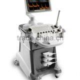
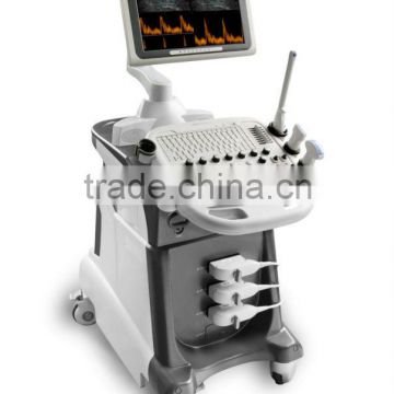
Abdominal/ Vascular/OB/GYN/4D Ultrasound ISpark 480 With CE Approved
Related Products
-
 AJ-6100B LCD Portable Pseudo Color US Ultrasound Scanner ChinaUS$ 100 - 1,000MOQ: 1 Piece
 -
ISpark 380 Ultrasonic Machine 3D Color DopplerUS$ 10,000 - 20,000MOQ: 1 Unit
-
4D Color Doppler With 4D Real Time Live Probe ISpark 480US$ 10,000 - 30,000MOQ: 1 Unit
-
Popular ISpark 380 High Performance Mature Technology Color Doppler Echo Machines With CEUS$ 10,000 - 20,000MOQ: 1 Unit
-
3 Channel Digital Electrocardiograph With CE ApprovedNegotiableMOQ: 1 Piece
Specifications
4D Color Doppler iSpark 4801.CE & FDA approved
2.3D/4D imaging; Strain Imaging; Free-hand 3D, 4D Real Time Live Probe
4D Color Doppler iSpark 480 with CE & FDA Approved
GENERAL DESCRIPTION
Color Doppler iSpark 480 is the fruit of innovative technologies, not only professionally support 3D/4D, Strain imaging and Wide field of view etc. high end functions, but also with 3D-PW, adaptive sound optimization, coded excitation and auto IMT etc innovative imaging processing technologies. Ergonomic design is devoting to facilitate exams in a wide variety of clinical environments. Comfort, convenience, scanning flexibility and efficiency is what iSpark 480 would like to provide to you.
FULL CLINICAL APPLICATION
3D/4D imaging;
Strain Imaging;
Free-hand 3D;
3D-PW;
Anatomic M Mode;
Trapezoid Imaging;
WFOV;
Color Flow Imaging;
CW;
Power/Directional Power Doppler;
Auto IMT Measurement;
TDI;
THI;
Real Time Triplex;
Clinic Applications
Abdominal, Vascular, OB/GYN, Cardiac, Urology, Breast, Small Parts, Musculoskeletal
Image Modes
B mode, M mode, Color Doppler (CFM), Doppler Spectrum Mode (PWD), Power Doppler Mode
Measurements & Calculations
OB : HR, Velocity(D), Accel, Auto Trace, Point Trace, RI, SI, M Trace(L), M Trace(H), Velocity(C), CRL, BPD, HC, AC, FL, OFD, HL, GS, Radius, Ulna, Tibia, Fibula, N.B.L, AFI…
Radiology: Peak Sys, Peak Dys, Mean, Time, HR, Velocity(D), Accel, Auto Trace, Point Trace, RI, SI, M Trace(L), M Trace(H), Flow Velocity, D Flow, T Flow, Lt. Kidney, Rt. Kidney, IMT…..
Cardiac: AR, LVOP, TR, PUL.VALVE, PUL.VEIN, RV, BF, LVET, Time, HR, Auto Trace, Point Trace, LAD/AOD, MPAD, RVEDd, RVEDs, LVM, BSA(Input), HR(Input)……
Vascular: VFD,VFA, Time, HR, Velocity(D), Accel, Auto Trace, Point Trace, RI, SI, M Trace(L), M Trace(H), Velocity(C), IMT, %DST, %AST, Dist, Area, Ellipse, Cross, Angle, Dist Ratio……
GYN: Uterine, Cervix, UT_L/CX_L, Lt.Ovary, Rt.Ovary, Endo L, DO Follicle, Uterus Body, Dist, Area , Ellipse, Cross, Angle, Dist Ratio, Para, M Trace(L), M Trace(H)…….
Small Part: HR, Accel, Velocity(D), Auto Trace, Point Trace, RI, SI, M Trace(L), M Trace(H), Dist, Rt.Thyroid, Lt.Thyroid, Area, Ellipse, Angle, Para, Dist Ratio, Diameter……
Urology: Lt.AG, Lt.AG, Lt.Testicle, Lt.Testicle, Lt.Sperm, Lt.Sperm, Rud.Urine, Prostate, Auto Trace, Point Trace, Accel, HR, RI, SI, Diameter, Joint Angle
IMAGE FEATURES
1. Display
Display Depth: up to 35cm,
Probe dependent: Convex: 18 steps, Linear: 14 steps
Display Gray Levels: 256; continuous variable contrast and frame rate up to 586/s
Display Format: B, 2B, 4B, M, B/D, B/M, B/C, B/C/MC, B/D, B/P, B/C/D
Image Orientation: Left/right B mode reversal and up/down image invert 90, 1800 rotation
Magnification: Zoom with pan capability in real-time or freeze B, C and M mode
Annotation: Allows the user to annotate anywhere on the image with pre-defined annotation list and anatomical body marks
Screen Display: Display of all patient and exam related imaging parameters, and on-screen documentation of image parameters in single / dual display modes
2. 3D Ultrasound
Offer freehand 3-D ultrasound with user training tools
Offer 3-D viewing and editing slices to remove unwanted tissue structure from arbitrary angles
Offer 3-D display in B-mode and Color Doppler simultaneously
3D Multi-gate Spectrum Doppler
3. 4D package(including Volume probe,4D software,4D hardware)
4. Image Review
CINE Review: Variable speed motion review and frame-by-frame review Storage only limited by internal memory of the system board
Standard: 1,024 B-Mode frame and 170 seconds M-Mode data
Standard: 520 color frames and 380 seconds Doppler Spectrum
Post Procession and measurements
5. Image Management
Storage only limited by hard disk of the system board
Standard: 160 GB hard disk drive for local image storing
CD-RW /DVD Drive as removable read, write, archive & storage, USB, S-Video, VGA
Standard: 2,000 Images w/ TIF format on CD
DICOM & PACs Compatible
standard CONFIGURATION
- iSpark 480 with Two active probe connectors
- 15 inch high resolution and contrast LCD
- 4D Real Time Volume Probe
- 4D package(including 4D software,4D hardware)
- 3.5 MHz Multi-frequency Convex probe
- 5120 system processing channels
- 177 dB dynamic range
- Display: B, C, M, CFM(Color Flow Mapping),PWD(Pulse Wave Doppler), PD(Power Doppler), DPI(Directional Power -- Dpower)Pulsed Wave Doppler (PW)
- Display mode: B, 2B, 4B, M, B/PWD, B/M, B/C, B/Power, C/Power, B/DPower, C/DPower, B/C/PWD
- Scanning Depth: 35cm
- Frame: 586 frame/s
- DICOM 3.0
- 160G Hard Disk
- DVD RW
- Remote system diagnosis and online service
STANDARD SPEICAL FUNCTIONS
- Coded Excitation
- Trapezoid Imaging
- Automatic Intimate media thickness (IMT) Measurement
- Tissue Harmonic Imaging(THI-phase)
- Spatial Compounding Imaging (SCI)
- Tissue Restoration Image (SRA+Structure enhancement)
- 2D Color flow estimation
- Color Zoom
- Synthetic Aperture
- Wide Field of View (WFOV) Imaging
- Automatic Sound Speed Optimization
- Crystal imaging
Optional
- Free Steering M software
- 3D Multi-gate Spectrum Doppler
- Elastic (Strain) Imaging
- 3D software(B mode)
- Cardiac function package(including TDI, Strain Rate Imaging, HPRF Imaging)
- DICOM ultrasound work station(including Main unit ,software, monitor)
- DICOM ultrasound work station(including software and monitor)
- Three probe sockets
OPTIONAL PROBES
- Linear probe
- Phased Array Probe
- Trans-vaginal Array Probe (180°)
Related Pictures:

Send Inquiry to This Supplier

You May Also Like
-
ULTRASOUND SCANNER/ESCÁNER DE ULTRASONIDONegotiableMOQ: 1 Piece
-
Best Selling Easy to Operate Low Noise Medical Infusion Pump With Large LCD DisplayUS$ 285 - 350MOQ: 1 Set
-
2000 Shots 2015 Ultrasonic Fat Reduction Hifu 0.1-2J Slimming Treatments Hifu Shape MachineNegotiableMOQ: 1 Set
-
Veterinary Hospital Scanner Ultrassom Sonoscape a5 VetNegotiableMOQ: 1 Set
-
Popular Min Beauty Equipment New Korean Technology Portable Cavitation Rf Slimming MachineUS$ 100 - 999MOQ: 1 Set
-
12.8V40Ah LiFePO4 for Mobile Medication CartsNegotiableMOQ: 50 Packs
-
Professional Original 3rd-generation System Quantum Physiotherapy Diagnostic Imaging Devices Factory PriceUS$ 1 - 120MOQ: 1 Set
-
One-armed Electrical Endoscopic Pendant (KL-T.IB J )NegotiableMOQ: 1 Set
-
China Manufacturer New Style Of Vein Finder With Low PriceUS$ 3,300 - 3,300MOQ: 1 Piece
-
China Supply Hotsale Link Digital Non-Interlaced Portable Ultrasound Scanner 200D With Low PriceUS$ 1,500 - 1,500MOQ: 1 Set


